In the world of chiropractic care, the use of x-rays plays a crucial role in accurately assessing and diagnosing patients. At Henry Chiropractic, owned and operated by Dr. Craig Henry, along with the expertise of Dr. Aaron Hixon, x-rays are recognized as an essential tool in providing the best possible care for patients in Pensacola, FL and the surrounding communities. With a commitment to improving health and wellness, the use of x-rays allows for a comprehensive understanding of each patient’s individual needs. Through a combination of chiropractic techniques and advanced diagnostic imaging, Henry Chiropractic utilizes x-rays to deliver effective and personalized treatment plans. Discover the importance of x-rays in chiropractic diagnosis and how it can improve your overall well-being.

Table of Contents
I. Introduction
Welcome to the world of chiropractic care! If you’ve ever experienced back pain, neck pain, or any other musculoskeletal issue, you might have considered visiting a chiropractor. Chiropractic care focuses on the alignment and function of the spine and nervous system to promote overall health and wellness. One of the key tools in chiropractic diagnosis is the use of X-rays. In this article, we will explore the importance of X-rays in chiropractic diagnosis and how they play a crucial role in guiding treatment planning and customization.

This image is property of sa1s3optim.patientpop.com.
II. What is Chiropractic Diagnosis?
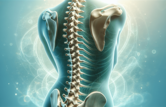
Chiropractic diagnosis is the process of identifying and understanding the root cause of pain and dysfunction in the musculoskeletal system. Chiropractors are skilled in assessing the spine, joints, and surrounding tissues to pinpoint areas of misalignment, nerve interference, and other issues that may be contributing to pain or decreased function. X-rays are an invaluable tool in this diagnostic process, providing chiropractors with detailed images of the spine and other structures to aid in accurate diagnoses and effective treatment planning.

This image is property of www.reinhardtchiropractic.com.
III. The Role of X-rays in Chiropractic Diagnosis
A. Understanding X-rays
X-rays, also known as radiographs, are a type of electromagnetic radiation that can penetrate the body and create images of internal structures. They are commonly used in medical imaging to diagnose various conditions and are an important tool in chiropractic care. X-rays allow chiropractors to visualize the bony structures of the spine, joints, and surrounding tissues, providing valuable insights into the alignment, function, and potential abnormalities that may be present.
B. Visualization of Spinal Structures
One of the primary roles of X-rays in chiropractic diagnosis is to provide a visual representation of the spinal structures. These images allow chiropractors to identify any misalignments, degenerative changes, fractures, or other structural abnormalities that may be contributing to pain or dysfunction. By accurately visualizing the spine, chiropractors can determine the best course of action to restore optimal function and alleviate discomfort.
C. Determining the Root Cause of Pain and Dysfunction
X-rays play a vital role in identifying the root cause of pain and dysfunction in the musculoskeletal system. By examining the alignment of the spinal vertebrae and assessing the relationship between the bones and surrounding soft tissues, chiropractors can identify areas of nerve compression, joint dysfunction, or other structural issues that may be causing symptoms. Identifying the underlying cause of the problem allows chiropractors to develop targeted treatment plans that address the specific issues and promote long-term healing.
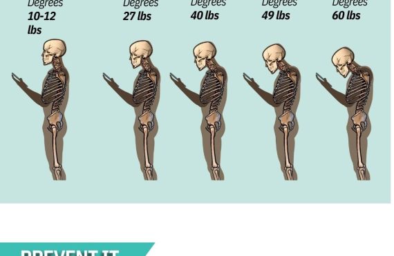
D. Assessing Spinal Alignment and Posture
Maintaining proper spinal alignment and posture is essential for optimal health and function. X-rays enable chiropractors to assess the alignment of the spinal vertebrae and identify any areas of misalignment or subluxation. By analyzing these images, chiropractors can determine the most effective course of action to restore proper alignment and posture. This may include spinal adjustments, exercises, and lifestyle modifications tailored to each individual patient.
E. Detecting Skeletal Abnormalities
In some cases, X-rays may reveal skeletal abnormalities that go beyond simple misalignments. These abnormalities may include congenital conditions, such as scoliosis or spinal deformities, or acquired conditions, such as fractures or tumors. Identifying these abnormalities through X-ray imaging allows chiropractors to make appropriate referrals and collaborate with other healthcare providers to ensure comprehensive and well-rounded care for their patients.
F. Identifying Joint Dysfunction
X-rays also provide valuable information about the health and function of the joints. By assessing the joint spaces, bone density, and any signs of degeneration or inflammation, chiropractors can gain insights into the overall joint health. This information allows them to develop appropriate treatment plans that target joint dysfunction and improve joint mobility, reducing pain and promoting optimal function.
G. Evaluating Soft Tissue Injuries
While X-rays primarily focus on visualizing bony structures, they can also provide some information about the surrounding soft tissues. Soft tissue injuries, such as muscle strains or ligament tears, often accompany spinal and joint issues. X-ray imaging can help chiropractors identify any signs of soft tissue damage, such as swelling, fluid accumulation, or calcifications. This information aids in developing comprehensive treatment plans that address both the bony and soft tissue components of musculoskeletal dysfunction.
H. Assessing Progress and Treatment Effectiveness
X-ray imaging is not only useful for initial diagnosis but also for tracking the progress of treatment and evaluating its effectiveness. By comparing follow-up X-rays to the initial images, chiropractors can objectively assess changes in spinal alignment, joint health, and overall structural improvements. This allows them to make adjustments to the treatment plan as needed and ensure that patients are progressing towards their desired outcomes.
I. Guiding Treatment Planning and Customization
X-rays provide chiropractors with a comprehensive view of the spine and related structures, which is crucial for developing personalized treatment plans. These images allow chiropractors to tailor their approach based on the individual patient’s specific needs, taking into account the severity and location of misalignments, the presence of degenerative changes, and other factors. This personalized approach ensures that each patient receives the most effective and appropriate care for their unique condition.
J. Patient Education and Informed Decision Making
X-ray images are invaluable tools for patient education and informed decision making. By visually illustrating the structural abnormalities and dysfunctions, chiropractors can empower their patients to understand the underlying causes of their symptoms. This knowledge enables patients to actively participate in their treatment plans, make informed decisions about their care, and take steps towards maintaining long-term spinal health.

This image is property of chrisbaileydc.com.
IV. The Process of X-ray Imaging in Chiropractic
A. Patient History and Examination
Prior to ordering X-rays, chiropractors conduct a thorough patient history and examination. This includes discussing the patient’s symptoms, medical history, and any previous imaging or interventions. Chiropractors also perform a physical examination, which may involve assessing posture, range of motion, and conducting orthopedic and neurological tests. This information helps chiropractors determine whether X-rays are necessary and which areas need to be imaged.
B. X-ray Ordering Process
Once the decision to proceed with X-ray imaging is made, chiropractors typically order X-rays through a radiology facility. They specify the desired views and any additional instructions based on their clinical findings during the examination. The radiology facility then schedules the patient for the imaging procedure, ensuring that safety measures are followed, such as protecting the patient from unnecessary radiation exposure.
C. Radiographic Positioning and Technique
During the X-ray imaging procedure, the patient is positioned according to the specific views requested by the chiropractor. This may involve standing, sitting, or lying on a table, depending on the area being imaged. The radiologic technologist ensures proper alignment and positioning to obtain clear and accurate images. X-ray machines emit a small amount of radiation, which passes through the body and creates an image on the film or digital sensor behind the patient.
D. Radiation Safety and Protection
Radiation safety is of utmost importance during X-ray imaging. Chiropractors and radiologic technologists follow strict protocols to ensure patient safety and minimize radiation exposure. This includes the proper use of lead aprons and thyroid shields to protect sensitive organs from radiation, as well as using the lowest possible radiation dose required to obtain diagnostic images. Additionally, pregnant women and children are given extra consideration, and alternative imaging methods may be used to avoid unnecessary exposure.
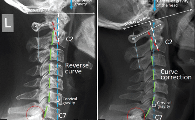
E. Image Analysis and Interpretation
Once the X-ray images are obtained, they are analyzed and interpreted by the chiropractor. This involves carefully evaluating the alignment of the spinal vertebrae, assessing joint spaces, identifying any degenerative changes, and noting any abnormalities or injuries. The chiropractor compares the images to their clinical findings and patient history to develop an accurate diagnosis and treatment plan. Clear communication and collaboration with the patient are essential during the interpretation process to ensure understanding and engagement in the treatment process.

This image is property of doc.vortala.com.
V. Potential Risks and Limitations of X-ray Usage
While X-rays are a valuable diagnostic tool, it is important to acknowledge their potential risks and limitations.
A. Radiation Exposure
X-rays involve exposure to ionizing radiation, albeit in small amounts. While the benefits of X-ray imaging generally outweigh the risks, it is crucial to minimize radiation exposure whenever possible. Chiropractors and radiologic technologists follow strict safety protocols to ensure patient safety, such as using lead shielding and employing appropriate techniques to reduce radiation doses.
B. Consideration for Pregnant Women and Children
Pregnant women and children are more sensitive to radiation and its potential effects. Chiropractors carefully assess the need for X-rays in these populations and weigh the benefits against the risks. Whenever possible, alternative imaging methods that do not involve ionizing radiation, such as ultrasound or MRI, may be recommended for pregnant women. For children, lower radiation doses and multiple precautions are taken to ensure minimal exposure.
C. Misinterpretation of Images
Interpreting X-ray images requires expertise and careful analysis. As with any diagnostic tool, there is a risk of misinterpretation. Chiropractors undergo extensive training to develop the skills necessary to accurately analyze X-ray images. However, collaboration and communication with other healthcare providers, as well as follow-up imaging or consultations, may be necessary to confirm diagnoses and ensure optimal care.
D. Overreliance on X-ray Findings
X-ray imaging is just one part of the comprehensive chiropractic diagnostic process. While X-ray images provide valuable information, they do not provide the complete picture of a patient’s condition. Chiropractors take into account the patient’s history, physical examination findings, and other imaging modalities, along with X-ray results, to form a comprehensive diagnosis and treatment plan. Overreliance on X-ray findings alone can lead to incomplete or inaccurate diagnoses.

This image is property of tebbyclinic.com.
VI. Conclusion
X-ray imaging plays a critical role in chiropractic diagnosis and treatment planning. It provides chiropractors with valuable insights into the alignment, function, and potential abnormalities of the spinal structures, facilitating accurate diagnoses and personalized treatment plans. While there are potential risks and limitations associated with X-ray usage, chiropractors follow strict safety protocols to minimize radiation exposure and maximize patient safety. As a patient, understanding the importance of X-rays in chiropractic diagnosis empowers you to actively participate in your treatment plan and make informed decisions about your health. If you are experiencing musculoskeletal pain or dysfunction, consider consulting with a qualified chiropractor, such as Dr. Craig Henry or Dr. Aaron Hixon of Henry Chiropractic in Pensacola, FL, who can utilize X-ray imaging to guide your personalized care and help you achieve optimal spinal health.